What The Research Says
Several recent studies have helped uncover brain changes in people with bipolar disorder.
In a 2019 study, researchers found that amygdala activation and connectivity in the brain are different between people with bipolar disorder and major depressive disorder. This finding may help doctors distinguish between the two conditions.
In a study from 2018, researchers looked at MRI scans and visualizations to determine the differences between people with bipolar disorder and people in control groups.
They found that when a person with bipolar disorder was neither in a state of mania nor a state of depression, their brains responded in the same way as the brains of the people in the control group.
However, the researchers did find brain changes in people in a state of mania or depression. In particular, their visual cortices changed appearance, showing less activity than the brains of those in the control group.
The study suggests that changes in sensory areas of the brain may also indicate bipolar disorder.
In a recent review , researchers found that evidence across several studies indicates widespread patterns of subcortical volume, lower cortical thickness, and disrupted white matter integrity. They suggest that future research may help determine key markers and help with the diagnosis of bipolar disorder.
Researchers also looked at how both the structure and the processes of the brain looked using MRI in
What Can An Eeg Find That An Mri Cant Find
Your brain works in wonderful and mysterious ways allowing you to walk, talk, and do everything that a human can do. Thats why we take so much care to protect our heads from injury.
But, what happens when the brain doesnt work the way its supposed to? The United Nations reported that about 1 in 6 people on earth today have some kind of neurological disorder!
Sometimes, doctors can intervene and remedy some of these brain disorders. But, they have to get a good look at your brain and how it works. Keep reading to learn the difference between an EEG and an MRI and how they help doctors understand your brain better.
Eeg And Quantitative Eeg
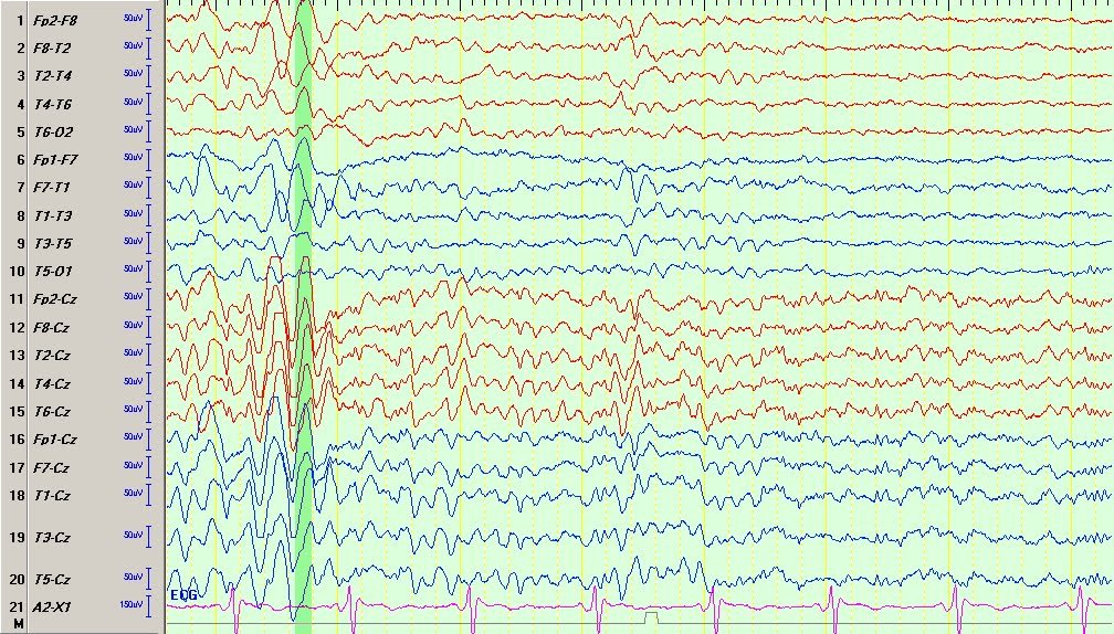
The EEG signal is composed of synchronised synaptic potentials in the cerebral cortex and appears as wave forms made up of different frequencies and rhythms. In the EEG of healthy adults and adults in a waking state, the activity consists mainly of alpha waves in the 813 Hz frequency range and some beta waves in the 1430 Hz frequency range, while there are few theta waves in the 47 Hz frequency range and almost no visible delta waves in the 0.53 Hz frequency range. Table 1 shows in which neural networks the different frequencies can be found . During drowsiness and sleep, an EEG will show more slow waves. Drugs that affect the brain can also change the speed of the EEG rhythms.
Table 1
|
EEG rhythm |
¹ Also involved in synchronisation between brain regions
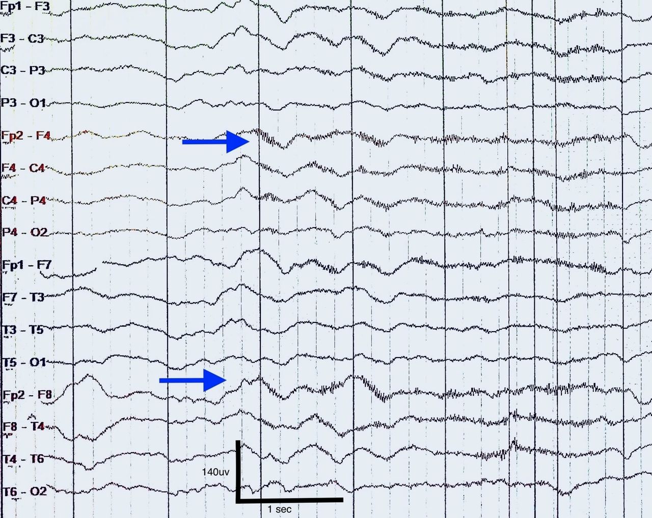
Epileptiform activity consists of sharp waves or a «spike-and-wave» pattern. This is a specific sign of epilepsy if the patient also has seizure symptoms which can fit the diagnosis. The probability of finding epileptiform activity in a patient with epilepsy increases if the EEG is recorded while the patient is sleeping .
Fig. 1
Recommended Reading: What To Do If Someone Is Having An Anxiety Attack
Demographics And Medical Characteristics
Analyses revealed age and gender effects on EEG power. Age was negatively correlated with Delta, Theta and Alpha2 power in the frontal region , but positively correlated with Beta1 and Beta2 power in the central region . There were also significant differences between genders in EEG power across all regions at Theta and both Alpha and Beta frequency ranges , with females displaying higher EEG power than men.
We conducted repeated measures ANOVAs to examine medication effects on EEG power. In BP, no significant effects of Medication were detected at any frequency. In SZ, there was a significant effect of Medication =6.25, p=.014) at Alpha2, with unmedicated patients exhibiting greater power than medicated patients. There was also a significant Region by Medication interaction =3.46, p=.024), driven by parietal and temporal regions only, whereby unmedicated patients showed greater power than patients on medication. At Beta1, there was a significant Region × Medication interaction =3.15, p=.034) that was driven by the parietal region only , with unmedicated patients displaying greater power. To account for these effects on our EEG variables, we included age, gender, and medication status as covariates in all subsequent analyses.
Resting Eeg Power And Coherence

Oscillations of electrical activity recorded by EEG reflect synchronized neuronal activity . Power within specified frequency bands indexes the average magnitude of oscillations over a specified time range, and coherence measures the extent of oscillatory coupling between two signals independent of their power. Both power and coherence of electrical activity during rest have been associated with fMRI measures of Default Mode Network functioning . The DMN is a resting state network that includes the precuneus, posterior cingulate cortex, medial prefrontal cortex and temporoparietal junction , and is generally more active at rest than during task performance . DMN is thought to be involved in self-referential thought and autobiographical memory retrieval , both of which are disturbed in psychotic disorders . While fMRI studies have characterized the spatial distribution of resting state networks, EEG measures offer unique information on the strength and synchronization of neuronal activity at high temporal frequencies.
Don’t Miss: What To Do To Stop A Panic Attack
What Part Of The Brain Is Responsible For Bipolar Disorder
A given brain hemisphere is responsible for either positive or negative events. The left hemisphere is responsible for the positive events and the right hemisphere is responsible for the negative events. Due to the reason that each hemisphere is responsible for certain types of events, every person has a dominant hemisphere. This hemisphere makes that person view the world in a certain way. A part of the brain is responsible for our mood. This part of the brain is called the limbic system. One of the parts is called the amygdala. The amygdala is an almond shaped structure in the temporal lobes responsible for decision making and emotional behavior. The amygdala is responsible for bipolar disorder and schizophrenia..
Diagnosis Can Be Complicated
If you have received a diagnosis or believe you might have Bipolar Disorder, formerly called manic depression, you probably know that getting a diagnosis is not a straightforward process. There is no blood test, for example, that definitively tells you that you have the illness. Mental health professionals use the DSM-V and take a survey of your symptoms and ask you how it impacts your life and relationships and uses that checklist to determine which type of Bipolar you may have. The Depression and Bipolar Support Alliance report that 7 out of 10 people are misdiagnosed at least once and that the average length of time from first experiencing symptoms and diagnosis is a decade. Every person is unique and your illness may not look exactly like it does for someone else. It can also change over time, becoming more or less severe.
Don’t Miss: How Is Bipolar Depression Different From Other Depression
When Do Doctors Recommend Brain Scans For Bipolar Disorder
In general, doctors do not recommend brain scans to diagnose bipolar disorder.
According to the Depression and Bipolar Support Alliance, there are currently two reasons that a doctor may order a brain scan for a person with bipolar disorder.
The first reason is to determine if a condition such as a stroke or a tumor could be causing a persons symptoms instead of bipolar disorder. However, this is rare.
The second reason is to be a part of research. Researchers are conducting studies to see whether or not there are identifiers within the brain that can help diagnose bipolar disorder. The results may help a doctor distinguish between bipolar disorder and major depressive disorder.
Ological Challenges And Limitations
One considerable challenge when reviewing the literature is the range of methodologies employed that result in difficulties comparing one study to another. Here, we outline the differences in participant selection, EEG recording and analysis that could impact the results reported in this review.
Study Size, Composition and Controls
The sample size of studies varies between n = 20 and n = 1,344 with three quarters of studies based on less than 100 participants. The median is 60 with similar numbers of controls and patients in the majority of studies. For most studies the age of participants were adults in the range of 25â45.
Interestingly most of the studies in this review were skewed towards male participants . This pattern is found for all disorders except for depression, bipolar disorder, panic disorder, anxiety and OCD . The largest gender disparity is seen for ADHD , schizophrenia , autism and addiction . In addition, it was more common to study all-male participant groups compared to all-female participants . In some instances, the ratio of males to females was intentionally designed to reflect the relative proportions of sufferers in the general population, but at other times was a reflection of participant availability, limiting the generalizability of these results, especially towards the female population.
Clinical Groups and Assessment
Recording Configuration
Processing of the Signal
Frequency Band Definition
Table 2. Summary of frequency band parameters.
Don’t Miss: What Kind Of Mental Disorder Is Schizophrenia
Psychiatric Disorders Increase The Risk Of Epilepsy
Patients with psychotic or affective disorders will have an increased risk of developing epilepsy . The causal connection is complex and probably includes neurobiological, psychosocial and/or iatrogenic mechanisms . A sub-group of patients with recurrent brief unstable depressive episodes often have an abnormal EEG , and have more frequent comorbid epilepsy than patients with classic depressive or bipolar disorders .
Fig. 2
In a retrospective observational study of clinical EEG in acute psychiatry from 2006, abnormal EEG was identified in 17 % of the patients . A little less than half had known epilepsy, but the EEG results changed the therapy in only 12 % . This study was retrospective and involved few EEG tests. In our experience, the real significance of EEG may be greater, however, because we now have better access to MRI scans and use more antiepileptic drugs in psychiatry. Even though most EEGs are normal, the results may be extremely important for those patients whose tests reveal findings. EEG should therefore be carried out on patients with conditions characterised by rapid changes in mood or behaviour, muscle cramps or other brief, stereotypical seizure-like symptoms.
Dont Let Brain Issues Weigh Heavy On Your Mind
Your doctor should explain what tests they think you need and the results from those tests. If you feel uncomfortable with your doctor, dont shy away from getting a second opinion!
When something isnt working in your brain, it makes life difficult in frustrating and unexpected ways. The sooner you address the issue, the sooner you can get back to living your life again!
We hope you enjoyed reading this article and that you learned the differences between an EEG and an MRI. If youve suffered from a brain injury or have other neurological symptoms, contact us today for a free consultation and well help you get your mind right.
Don’t Miss: How To Deal With Dog Anxiety
Reasons A Doctor Would Recommend Tests For Your Brain
A complex organ like the brain can have any number of problems, even without injury. The Brain Foundation lists infections, autoimmune disease, seizures, and dementia among the most common neurological conditions doctors see.
The most common brain condition that leads to permanent disability in adults is ischemic stroke. This happens when the blood vessels get blocked, which restricts blood flow to the brain.
Also on that list is a traumatic brain injury. Even if the blow to your head doesnt break the skin, the brain can suffer enough damage to impair its function. Doctors can use brain imaging to find these injuries and decide if you need treatment or if treatment is even possible.
Eeg / Meg Based Diagnosis For Psychiatric Disorders: Volume Ii

-
Views
Keywords:EEG, MEG, Artificial intelligence, Machine learning, Diagnosis, Depression, Anxiety, Schizophrenia, Psychiatric disorders
Important Note: All contributions to this Research Topic must be within the scope of the section and journal to which they are submitted, as defined in their mission statements. Frontiers reserves the right to guide an out-of-scope manuscript to a more suitable section or journal at any stage of peer review.
You May Like: What Was The First Drug Used To Treat Schizophrenia
Best Way To Prepare For Electroencephalography
The Following Steps Should be Taken Before the Test:
- Wash your hair the night before the electroencephalography, and dont put any items in your hair upon the arrival of the test.
- Inquire as to whether you should quit taking any drugs before the test. You ought to likewise make a rundown of your meds and offer it to the professional playing out the electroencephalography.
- Abstain from eating or drinking anything containing caffeine for at any rate eight hours before the test.
- Your PCP may request that you rest as little as conceivable the night before the test on the off chance that you should rest during the electroencephalography. You may likewise be given a narcotic to assist you with unwinding and rest before the test starts.
The electroencephalography can last 2 hours after the test, but you will still feel drowsy for a short time afterward if you had been given a sedative. This means that after the test you will need to be taken home by a responsible adult. You must stay out of the roads until the prescription dies down.
What Is Bipolar Eeg
Bipolar EEG is a type of EEG that records the electrical activity of the brain from two symmetrical positions on both sides of the head. It is also known as a scalp EEG. It is used to diagnose and monitor changes in activity of the brain in subjects who are suspected of having seizures or other neurological disorders. Bipolar EEG recording creates a waveform similar to the letter E in a graphic display. This waveform is known as a correlogram. The shape of the wave depends on the wave form of the EEG, but there are some specific waveforms that correspond to specific types of brain wave activity..
Also Check: Does Depression Run In Families
What Is Driver Attention On Honda Accord
- harry limThe Feature: The CR-V was the first Honda to come with a Driver Attention Monitor. This feature uses an angle sensor to measure the degree of steering-wheel corrections by the driver to maintain a proper lane position. If it senses too much correction activity, it will notify the driver to take a break.
Potential Mechanisms Of Lithium
Influences on neurotransmitters, signaling cascades, neurotrophic factors, and neuroplasticity cascades are mechanisms of action for lithium.67 Mechanisms that modify integrative brain function with lithium might be involved in most of the mentioned mechanisms, and the relevance of these mechanisms with EEG changes will be discussed in the following sections.
Don’t Miss: How To Treat Binge Eating Disorder
Delta/alpha Frequency Activity Group Differences
During REC differences were found for delta/alpha frequency activity across all electrodes: F3 F4 C3 C4 P3 P4 . Where SCZ and MPD delta/alpha frequency activity was higher than CON . Then MPD delta/alpha frequency activity was higher compared to BPD for right hemisphere only .
During REO differences were found for delta/alpha frequency activity across all electrodes, except for F3: F4 C3 C4 P3 and P4 . Where SCZ, BPD and MPD delta/alpha frequency activity was higher than CON .
During the CPT difference were found for delta/alpha frequency activity across all electrodes, except for F3: F4 C3 C4 P3 P4 . BPD and MPD delta/alpha frequency activity was higher than CON .
There Is Another Choice Neurofeedback Training
Drugs act on the body`s chemistry in an attempt to control the symptoms of bipolar disorder. The source of the problem, improper brainwave balances, is not addressed at all. Neurofeedback training is different in that it goes to the source of the mood swings, and helps correct brainwave functions in the brain itself. Once the brainwaves are more balanced, the symptoms can disappear on their own.
There are many studies showing that neurofeedback can be an effective treatment for Bipolar Disorder and other mood disorders like depression, anxiety, and PTSD.
You May Like: Which Of These Is Associated With Binge Eating Disorder
When To Ask For An Eeg
It is common sense to consider EEG under certain specific circumstances, such as those listed in Box 3. This list is neither prescriptive nor exhaustive, but it implies that clinicians should be highly vigilant for possible organic factors underlying any psychiatric presentation.
BOX 3 Some situations in which an EEG might be requested
An EEG might be requested:
to interpret paroxysmal activity
to analyse sleep disorders
to establish the cause of cognitive decline when there is a suspicion of Huntington’s disease, CreutzfeldtJakob disease, chronic delirium, etc.
A serial, ambulatory or video EEG may be useful in:
interpreting periodic behavioural dysfunction
Differential diagnoses under consideration
What Is An Mri

An MRI gives doctors a map of the brain structures. This structural information is often used to determine how certain brain areas compare with other normal brains to look for abnormal structures like a tumor.
An MRI wont show any brain activity though, so many doctors will recommend other brain imaging methods first. Its more expensive and harder to do an MRI test so its used only if the doctor thinks theres an abnormal growth.
Recommended Reading: How To Get Rid Of Relationship Anxiety