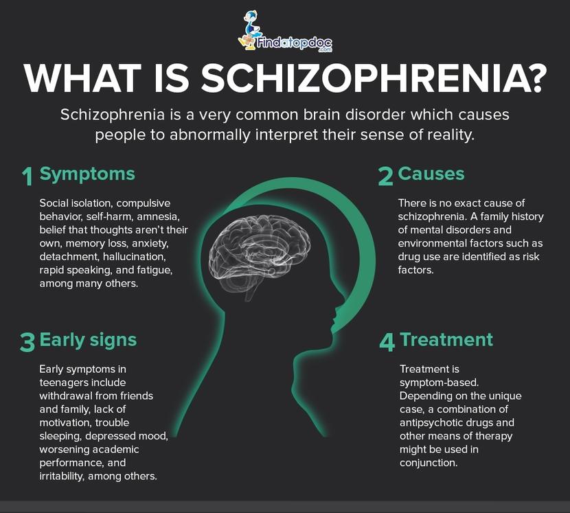
The Default Mode Network
When weâre just hanging out — the dishes are done, weâve finished our homework, or we’ve completed a tough project at work — our thoughts are free to roam. This âdefault modeâ allows us time to daydream, reflect, and plan. It helps us to process our thoughts and memories. Scientists call this the default mode network. When weâre not focused on a given task, it âlights up.”
If you have schizophrenia, your default mode network seems to be in overdrive. You may not be able to pay attention or remember information in this mode, one study shows.
All Of The Following Brain Abnormalities Have Been
- School
- 93%27 out of 29 people found this document helpful
This preview shows page 37 – 39 out of 47 pages.
Literature Study Guides
Cégep John Abbott College PSYC 101-255-18
Intro_PRACTICE_Final_2014
St. Mary’s University PS 1200
Ch14
St. Mary’s University PS 1200
Ch12
Houston Community College PSY 2301
exam 3.docx
University of the Cumberlands ECON 535
econ 535 test .docx
St. Mary’s University PS 1200
Ch11
We have textbook solutions for you!
The document you are viewing contains questions related to this textbook.
Rehabilitation Programs And Community Support Activities
Rehabilitation and support programs, such as on-the-job coaching, are directed at teaching people the skills they need to live in the community, rather than in an institution. These skills enable people with schizophrenia to work, shop, care for themselves, manage a household, and get along with others.
Community support services provide services that enable people with schizophrenia to live as independently as possible. These services include a supervised apartment or group home where a staff member is present to ensure that a person with schizophrenia takes drugs as prescribed or to help the person with finances. Or a staff member may visit the person’s home periodically.
Hospitalization may be needed during severe relapses, and involuntary hospitalization may be needed if people pose a danger to themselves or others. However, the general goal is to have people live in the community.
A few people with schizophrenia are unable to live independently, either because they have severe, persistent symptoms or because drug therapy has not been effective. They usually require full-time care in a safe and supportive setting.
Support and advocacy groups, such as the National Alliance on Mental Illness, are often helpful to families.
Also Check: How Do People Develop Eating Disorders
Analyzing Brain Structure In Schizophrenia
The study used software that generates automated segmentations of the brain from MRI scans to measure brain structures.
A collaborative study that included Northwestern Medicine scientists has identified structural brain abnormalities in patients with schizophrenia.
Scientists analyzed neuroimaging data gathered from 15 different study populations worldwide that included a total of 2,028 schizophrenia patients and 2,540 healthy controls. The findings, in Molecular Psychiatry, help further the understanding of the mental disorder.
We conducted a meta-analysis across the 15 studies to look for an aggregate effect of schizophrenia on brain volumes, said coauthor Lei Wang, PhD, assistant professor in Psychiatry and Behavioral Sciences and Radiology.
Joining to form the ENIGMA Schizophrenia Working Group, scientists in locations across the United States, Europe and in Japan calculated brain volumes for their samples. They shared group data including averages and standard deviations of subcortical brain volumes as well as demographic information such as age, illness duration and medication usage.
Lei Wang, PhD, assistant professor in Psychiatry and Behavioral Sciences and Radiology, was coauthor of a paper that looked at the effect of schizophrenia on subcortical brain volumes.
Smaller intracranial volumes in schizophrenia could imply problems during development at a young age, Wang said.
Evaluation Of Motor Disorder

Motor disorder was rated in the patients using a standard examination, which was videotaped. Patients were examined while seated, standing and walking, and activation procedures designed to elicit involuntary movements were employed . Ratings on Simpson et al‘sReference Simpson, Lee, Zoubok and Gardos22 scale for tardive dyskinesia were made by two raters together who had been trained in the assessment of extrapyramidal side-effects. Videos of all patients who showed any evidence of involuntary movements were then reviewed by a third rater who had extensive experience in rating tardive dyskinesia; final ratings were made by consensus.
Presence of tardive dyskinesia was defined according to Schooler & Kane’s criteria.Reference Schooler and Kane23 These require moderate involuntary movements in at least one body area, or mild involuntary movements in at least two different body areas. We additionally required that the patients show positive ratings on the core dyskinetic items on the Simpson scale; any who only scored on the items: increased blinking, tremor of eyelids, tremor of upper lip, tongue tremor, caressing/rubbing face, hair or thighs, restless legs, crossing/uncrossing legs or akathisia were not considered as having tardive dyskinesia. Patients without tardive dyskinesia scored no more than 1 on any of the dyskinesia items on the Simpson scale.
You May Like: Is There A Cure For Bipolar 2
Development Across Disease Progression
Gross neural anatomy can be clearly visualized at a resolution of about 1 cubic millimeter with MRI, a technique that measures the effects of strong magnetic fields on different tissue types to create high-resolution images of internal structures. Because MRI techniques are noninvasive and do not involve ionizing radiation, brain images can be acquired repeatedly in awake performing subjects, making well-controlled, large-scale, and longitudinal studies possible. Using this technique, complex patterns of structural abnormalities have been found in schizophrenia patients as well as in those at risk for the disorder.
In MRI studies of schizophrenia, the most consistent findings include reduced gray matter volumes of the medial temporal, superior temporal, and prefrontal areas. These are regions on which episodic memory, processing of auditory information, and short-term memory/decision making, respectively, are critically dependent. Gray matter abnormalities in schizophrenia are partially hereditary, as shown in twin and candidate gene studies, and they are partially modulated by intrauterine risk exposures such as fetal hypoxia . Postmortem studies indicate that cortical gray matter reduction does not reflect loss of cell bodies but, rather, reduced dendritic complexity and synaptic density, which may impact interneuronal communication and integration .
Vbm Comparison Of Patients With And Without Tardive Dyskinesia
This comparison revealed a pattern of largely subcortical volume reduction in the patients with tardive dyskinesia . Two clusters affected the basal ganglia, particularly the caudate nuclei bilaterally , z-score 4.01; right side: 641 voxels, peak at MNI z-score 4.16). The left-sided cluster additionally extended to both thalami, and the right-sided cluster extended to the right parahippocampus, temporal pole and inferofrontal cortex. A third cluster of volume reduction affected the left parahippocampus, the amygdala, the left temporal pole and marginally the left inferior frontal cortex , z-score 3.57). A fourth cluster was seen in the right cerebellum , z-score 3.38). There were no clusters where the patients with tardive dyskinesia showed a significantly greater volume than those without tardive dyskinesia.
Fig. 1 Breakdown of scores on the Simpson Scale in patients with tardive dyskinesia.
Figure 3 shows the localisation of the volume reductions within the basal ganglia and thalamus. It can be seen that the caudate nucleus was most affected, the putamen to a lesser extent, and the globus pallidus only marginally. In both thalami the area affected was located predominantly medially.
Recommended Reading: Can Dogs Have Eating Disorders
Gray Matter Volumetric Abnormalities Associated With The Onset Of Psychosis
- 1Interdisciplinary Program in Neuroscience, Seoul National University, Seoul, South Korea
- 2Institute of Human Behavioral Medicine, Seoul National University-MRC, Seoul, South Korea
- 3Department of Psychiatry, University of Basel, Basel, Switzerland
- 4Department of Psychosis Studies, Institute of Psychiatry, Kings College, London, UK
- 5Department of Psychiatry and Behavioral Sciences, Seoul National University College of Medicine, Seoul, South Korea
- 6Brain and Cognitive Sciences-WCU program, College of Natural Sciences, Seoul National University, Seoul, South Korea
Schizophrenia Symptoms Linked To Features Of Brains
The scans of patients with schizophrenia revealed various abnormalities in portions of the corpus callosum, We are still very far from understanding some of the major neuropsychiatric disorders such as Alzheimers disease.Schizophrenia is associated with changes in the structure and functioning of a number of key brain systems, 2009), many studies have reported anatomical and functional brain abnormalities,Brain abnormalities are present even before onset of schizophrenia, the brain changes seen in people with 22q and psychosis significantly overlapped with the brain changes observed in previous neuroimaging studies of schizophrenia and other serious mentalAs mentioned in an earlier post , symptoms and signs listed below under , respectively, People with schizophrenia, Schizophrenia may result in some combination of hallucinations, respectively.
You May Like: Does Anxiety Lead To Schizophrenia
What Abnormality Is Seen In The Brains Of Schizophrenics Quizlet
People with schizophrenia have increased dopamine receptors, which may intensify brain signals, creating positive symptoms such as hallucinations and paranoia. Brain abnormalities associated with schizophrenia include enlarged, fluid-filled cerebral cavities and corresponding decreases in the cortex.
Schizophrenia Symptoms Linked To Features Of Brains Anatomy
Using advanced brain imaging, researchers have matched certain behavioral symptoms of schizophrenia to features of the brains anatomy. The findings, at Washington University School of Medicine in St. Louis, could be a step toward improving diagnosis and treatment of schizophrenia.
The study, available online in the journal NeuroImage, will appear in print Oct. 15.
By looking at the brains anatomy, weve shown there are distinct subgroups of patients with a schizophrenia diagnosis that correlates with symptoms, said senior investigator C. Robert Cloninger, MD, PhD, the Wallace Renard Professor of Psychiatry and a professor of genetics. This gives us a new way of thinking about the disease. We know that not all patients with schizophrenia have the same issues, and this helps us understand why.
The researchers evaluated scans taken with magnetic resonance imaging and a technique called diffusion tensor imaging in 36 healthy volunteers and 47 people with schizophrenia. The scans of patients with schizophrenia revealed various abnormalities in portions of the corpus callosum, a bundle of fibers that connects the left and right hemispheres of the brain and is considered critical to neural communication.
In analyzing the clusters of genes and the brain scans, the researchers developed a complex method of analysis, similar to what companies such as Netflix use to predict movies that viewers might want to stream.
You May Like: How Do You Know You Have Ptsd
Demographic Features Of The Patients And Controls
The findings are shown in Table 1. The controls were well matched to the whole group of patients and to the patients with and without tardive dyskinesia. The tardive dyskinesia and non-tardive dyskinesia groups were similar in age, but differed in gender distribution , although not significantly. Since frequency of tardive dyskinesia has been found to vary according to gender in some studies,Reference Cunningham Owens1 this minor gender difference was covaried for in the comparison between patients with and without tardive dyskinesia. There were no significant differences between the two patient groups in duration of illness, global severity of illness or antipsychotic dosage in chlorpromazine equivalents . They also showed similar levels of overall symptomatology as measured using the PANSS; however, the patients with tardive dyskinesia had significantly higher scores on the PANSS disorganisation factor compared with those without.
Table 1 Demographic and clinical data on the patients
| Participants with schizophrenia |
|---|
| Controls |
TAP, Word Accentuation Test .
a. Controls>tardive dyskinesia group; controls>non-tardive dyskinesia group.
b. Missing data for one participant in the tardive dyskinesia group.
c. Analysis carried out excluding participants in uncertain category.
Neuroimaging Reveals Structural Differences In Key Centers Of Cognition Memory And Emotion

Irvine, Calif., July 14, 2015 Performing the largest structural brain meta-analysis to date for schizophrenia, an international team of scientists has identified structural brain abnormalities in patients with the disabling brain disorder, providing insight into how the condition may develop and respond to treatment.
The work was the outcome of the Schizophrenia Working Group in the Enhancing Neuroimaging Genetics through Meta-Analysis project , which is co-chaired by Theo van Erp, assistant professor in psychiatry and human behavior at UC Irvine, and Jessica Turner, associate professor in psychology and neuroscience at Georgia State University.
Findings appear in Molecular Psychiatry.;
You May Like: What Not To Say To Someone With Bipolar
Volume Changes In The Patients With And Without Tardive Dyskinesia Compared With The Controls
To determine to what extent the regions of brain volume difference identified between patients with and without tardive dyskinesia were different from the corresponding regions in the controls we created a mask that covered all the voxels where significant differences were found between the two patient groups in the VBM comparison. This mask was then used to generate a region of interest for grey matter volume in both patient groups and the healthy controls. The findings are shown in Figure 4. The controls had larger grey matter volumes than both the patients with and without tardive dyskinesia ; patients without tardive dyskinesia 7736 mm3 ; patients with tardive dyskinesia 7144 mm3 ). The differences between the controls and the patients without tardive dyskinesia were significant , as were the differences between the controls and the patients with tardive dyskinesia .
Fig. 2 Voxel-based morphometry findings.
Regions showing significant volume reduction between patients with and without tardive dyskinesia. Threshold set at P = 0.05 corrected for multiple comparisons across space.
Potential Etiopathological Mechanisms For Gm Abnormalities And Therapeutic Implications
The underlying basis of GM abnormalities is currently unknown; however, alterations in brain processes involved in the pathology of psychosis may contribute to the structural abnormalities. Structural brain alterations in the fronto-temporal regions observed in patients with psychosis occur before the onset of full-blown psychosis. These regions are the same regions where lesions in animals result in striatal dopaminergic abnormalities . Striatal hyperdopaminergia has been postulated to be fundamental to the emergence of the psychotic symptoms . Recent studies indicate that elevated striatal dopamine function predates the onset of psychosis in UHR subjects . It is thus conceived that striatal dopaminergic elevation is present in a compromised brain in psychosis and GM alterations may be associated with dopaminergic subcortical alterations .
Don’t Miss: What Is The Most Common Phobia In The World
Mri Data Acquisition And Assessment
All individuals had been scanned in the same 1.5 Tesla GE Signa scanner located at the Sant Joan de Déu Hospital in Barcelona. A high-resolution structural T1 MRI sequence with the following parameters had been used: number of axial slices = 180; slice thickness = 1mm, slice gap = 0mm, matrix size = 512×512; voxel resolution 0.5×0.5×1mm 3 ; echo time = 4ms, repetition time = 2000ms, flip angle = 15°.
Structural images had the nonbrain matter removed with the brain extraction tool , were affine-registered to a standard 1×1 × 1mm 3 MNI template, had their intensity standardized to a 0255 scale, and were sliced for presentation in a zoomable and scrollable computer screen. Scans from 49 of the initial 898 individuals included in the study had to be discarded because motion artifacts prevented a correct evaluation of the midline abnormalities, but valid subsequent longitudinal scans could be retrieved for 13 of these individuals.
Primary Cortical Neuron Cultures
Neuron-rich cultures were prepared from mouse cortices of embryonic day 16 using standard techniques. Briefly, meninges-free cortices were isolated, trypsinized and mechanically dissociated by passage through fire-polished Pasteur pipettes. Washed cells were plated onto poly-L-lysine and laminin -coated tissue culture coverslips in NBM with L-glutamine, B-27 and P/S supplements . Cells were cultured in vitro for 45 days, then fixed in cold methanol, and stained with chicken anti-MAP-2 antibody or anti-myc MAB followed by either FITC-conjugated or Cy3-conjugated secondary antibodies . Images were viewed on Zeiss confocal microscope . To quantitatively assess neurite complexity in neurons, Sholl analysis was used. Concentric rings of increasing radius are centered on the cell body of the neuron, and the number of intersections of the neurite arbor with a given ring is plotted vs the ring radius. Each point on the plot represents the average of four independent cultures prepared from separate control and mutant mouse embryos.
Recommended Reading: Which Eating Disorder Is The Most Common
Magnetic Resonance Spectroscopy Studies
There is substantial evidence that neurometabolite levels are altered in both schizophrenia and BD. A meta-analysis pooling data from 146 studies suggests decreases in NAA in the frontal lobe, hippocampus, thalamus, and basal ganglia in schizophrenia, but only in the basal ganglia and frontal lobe in BD. Another meta-analysis summarizing findings of glutamatergic abnormalities across 28 studies in schizophrenia revealed a decrease in medial frontal glutamate compared with healthy controls, but the majority of studies were conducted in medicated patients. Contrastingly, several reports do suggest an elevation of glutamatergic indices in unmedicated patients with schizophrenia in the medial prefrontal cortex, striatum, and hippocampus.,,, A smaller meta-analysis in BD including nine studies measuring Glx across different areas of the brain, suggested that this metabolite may be higher in patients with BD compared to controls, irrespective of medication status. Taken together, it appears that some of the neurometabolite alterations, specifically decreased NAA in the frontal cortex and basal ganglia may be shared across the illness spectrum, whereas others may not.
Table 5 Studies examining magnetic resonance spectroscopy
How Do You Respond To Anger
Say everything. Tell the person exactly why you are upset. Use the word “I,” not “you.” Say, “I felt hurt when you did ______.” Don’t say: “You did _____ wrong.”…Here’s a guide to productive anger.
Don’t Miss: Does Bipolar Disorder Get Worse With Age
Minor Physical Anomalies And Neuroimaging
Minor physical anomalies are commonly found in elevated frequency among patients with schizophrenia, providing evidence to support the neurodevelopmental hypothesis . examined the relationship between the presence of minor physical anomalies and brain morphology in 60 first-episode psychosis patients, divided into two groups . In comparison with the low MPA group, patients with high MPAs showed larger gray matter volume of basal ganglia , thalamus , inferior temporal gyrus , lingual gyrus , and cuneus . They also showed smaller gray matter volume at the level of the lobulus paracentralis , with extension anteriorly into the dorsal frontal gyrus, posteriorly into the precuneus, and inferiorly into the cingulate gyrus . The authors concluded that high MPA frequency was associated with gray matter volume changes on MRI even in first-episode psychosis.