The Tertiary Rod Pathway: Connections Between Rods And Certain Off Cone Bipolar Cells
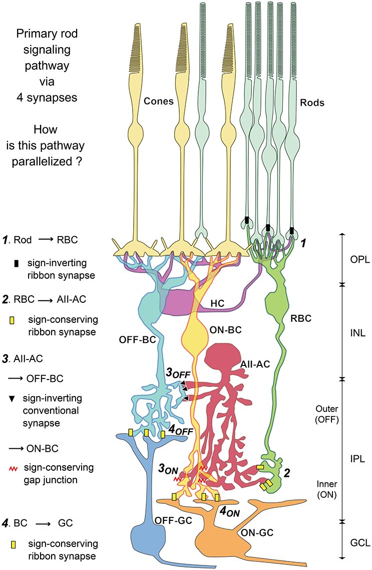
There is strong evidence for the connection between rods and certain OFF cone bipolar cells, as first proposed by Soucy et al. and Li et al. . Specifically, OFF bipolar types 3A, 3B, and 4 all make basal contacts with rod spherules in the mouse retina . These contacts coincide with excitatory glutamate receptors of the AMPA/kainate subtype providing a sign-conserving input consistent with the physiological responses of OFF cone bipolar cells . In addition, the direct connection between rods and certain OFF cone bipolar cells was demonstrated by paired recording in the ground squirrel retina . Direct rod input to OFF cone bipolar cells constitutes the tertiary rod pathway in the mammalian retina. Although the anatomical physiological basis for the tertiary rod OFF pathway is convincing, the function of this pathway is largely unknown, although it is thought to operate in the high scotopic/mesopic range.
Preparation Of Isolated Retina
All procedures conducted were approved by the Institutional Animal Welfare Committee. Adult New Zealand albino rabbits of either sex were used for this study. Rabbits were deeply anesthetized with urethane . Immediately prior to enucleation 2% lidocaine hydrochloride drops were applied topically to each eye. Retinas were isolated from the eyecup while immersed in carboxygenated Ames medium and mounted on 0.8 m black filter paper. Retinal cells were pre-labeled with 4,6-diamino-2-pheynylindole by incubating retinal pieces in Ames medium containing 5 M DAPI for 15 min.
Analysis Of Responses To Spot Stimuli
During the experiment, we first estimated the location over which subsequent spot stimuli were presented . To do so, we shifted a contrast-reversing spot, whose diameter could be manually increased or decreased, over the screen, until a position and diameter size were found that maximally stimulated the cell . Alternatively, we applied online analysis of responses to spatiotemporal white noise to obtain the receptive field parameters .
Estimation of optimal spot size
To estimate the spot size that best stimulated a cells receptive field center for further analyses, we presented spots of black and white contrast steps at 11 or 13 fixed diameters in random order, centered over the online-determined location. The spots were presented for 0.5 s on a background of mean light intensity and separated by 1 s of background illumination. Each spot size was presented on average 8 times. For each trial, we subtracted the average membrane potential measured over the 200 ms prior to the spot, and for each spot size, we averaged the baseline-subtracted responses over trials. For 4 cells, we presented the spots without an interval of background illumination between individual spots and only 6 spot diameters . These spots contrast-reversed at 1 Hz for 4 s, with different spot sizes separated by background illumination of 4 s, which was used for computing the baseline membrane potential.
Hyperpolarization index
Sustained-transient index
Latency
Center-surround index
Also Check: Depression Topography
Aii Amacrine Cell Fills
For technical reasons, including the targeting and/or identification of specific cone bipolar cell types, we did not think it was practical to address the photoreceptor connectivity of cone bipolar cells by individually filling cone bipolar cells. Therefore, we chose to adopt an alternative method, by filling AII amacrine cells. Because AII amacrine cells are coupled to most ON cone bipolar cells, this yields a bulk population of ON cone bipolar cells filled with Neurobiotin via the gap junctions with AII amacrine cells in the IPL, as first documented by David Vaney , reflecting the gap junction connections reported in early anatomical studies .
Nonlinear Inputs To Bipolar Cells

Similar to the contrast representation, the signal integration in bipolar cells is viewed as linear because of linear photoreceptor responses and of the ribbon synapses at the photoreceptor terminals . Support for linear signal integration comes from a small number of sample recordings in salamander bipolar cells that documented accurate response predictions to jittering gratings with a linear-receptive-field model as well as from observed response nulling in the glutamate release of mouse bipolar cell terminals under large reversing-gratings . Note, though, that the latter study focused on bipolar cells connected to alpha retinal ganglion cells whose major inputs likely come from few specific bipolar cell types , leaving open the question whether other bipolar cells in the mouse retina may differ in this respect. On the other hand, species-specific features might influence the observed nonlinearities. For example, the salamander retina has more photoreceptor types and the bipolar cells morphology might be more diverse compared to the mouse retina .
Don’t Miss: Mdd Vs Bpd
Contrast Processing By On And Off Bipolar Cells
Published online by Cambridge University Press: 19 November 2010
- DWIGHT A. BURKHARDT*
- Affiliation:Department of Psychology, University of Minnesota, Minneapolis, MinnesotaGraduate Program in Neuroscience, University of Minnesota, Minneapolis, Minnesota
- *
- *Address correspondence and reprint requests to: Dwight A. Burkhardt, University of Minnesota, n218 Elliott Hall, 75 E. River Road, Minneapolis, MN 55455. E-mail:
What Happens To Bipolar Cells When Light Hits The Retina
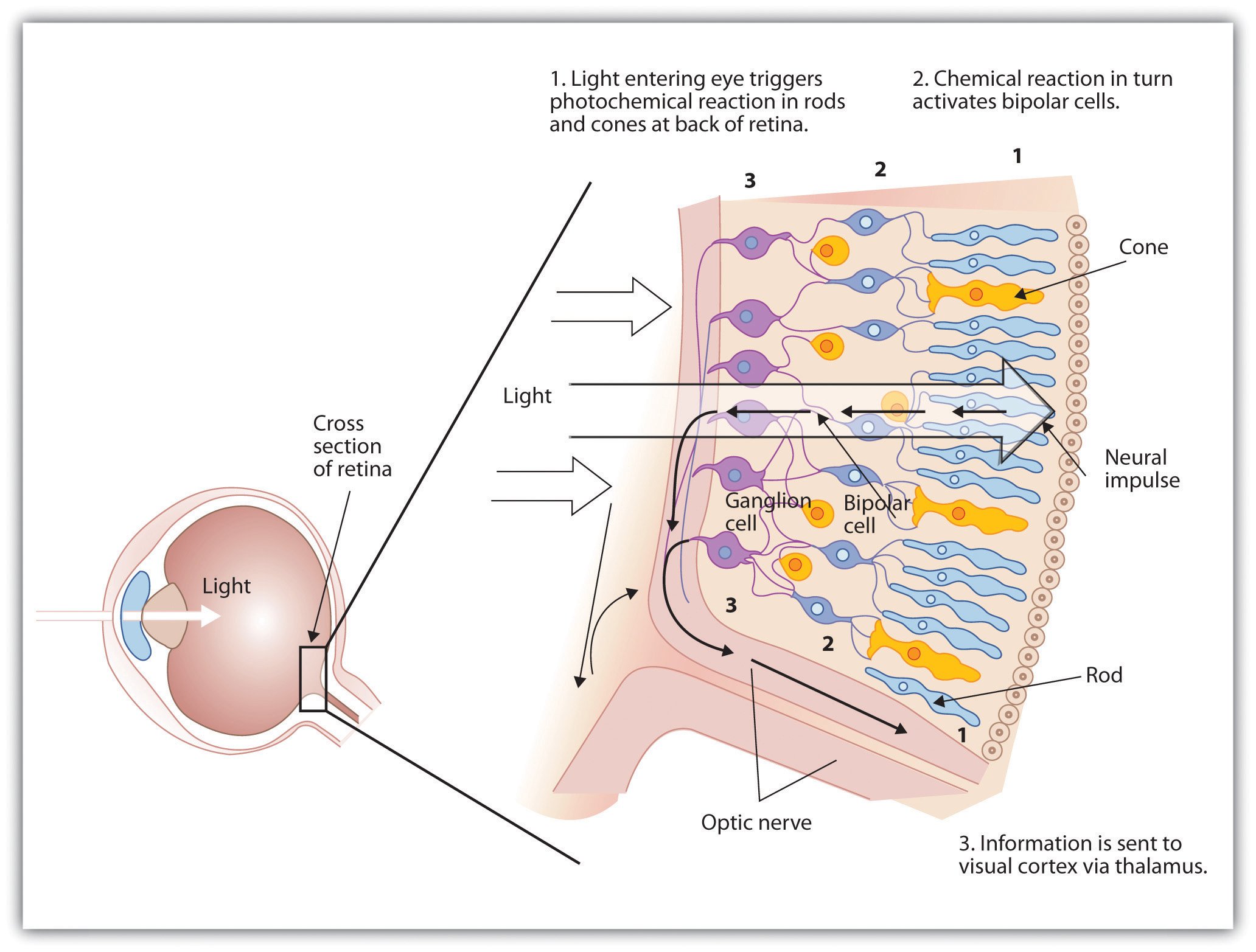
The visual pathway in the retina consists of a chain of different nerve cells. Light first travels through all the layers until it reaches the photoreceptor layer, the rod and cone layer. Bipolar cells can either hyperpolarize or depolarize with light, and they pass their signal on to amacrine cells or ganglion cells.
Don’t Miss: Fobia Meaning
What Are On And Off Bipolar Cells
bipolar cellsbipolar cellsOFFbipolar cellscells
. In respect to this, what are on and off cells?
The major functional subdivision of ganglion cells in the mammalian retina is into ON- and OFF-center ganglion cells. ON-center cells are depolarized by illumination of their receptive field center , while OFF-center cells are depolarized by decreased illumination of their RFC.
Likewise, what do bipolar cells release? Light responses in bipolar cells are initiated by synapses with photoreceptors. Photoreceptors release only one neurotransmitter, glutamate yet bipolar cells react to this stimulus with two different responses, ON-center and OFF-center .
People also ask, what are bipolar cells?
Bipolar cells are the central neurons of the retina which carry light-elicited signals from photoreceptors and horizontal cells in the outer retina to amacrine cells and ganglion cells in the inner retina. From: Encyclopedia of Neuroscience, 2009.
How many bipolar cells are in the retina?
Bipolar cell identification and diversity.There are more than ten types of bipolar cells in the mammalian retina. These typically consist of slightly more ON than OFF types plus a single type of rod bipolar cell (Box 1 Fig.
Spatiotemporal White Noise Analysis
Receptive field estimation
We visually stimulated the retina with binary spatiotemporal white noise in a checkerboard layout, where each square had a size of 30 µm and was updated randomly to black or white at 30 , 15 , 10 , or 7.5 Hz . For cells for which we also recorded natural movies, the squares had a size of 22.5 µm and were updated at 25 Hz or 12.5 Hz to fit the spatial and temporal resolution of the natural movies . Recording duration under spatiotemporal white noise was 23 ± 19 minutes . To remove slow fluctuations in the responses, the voltage traces were first de-trended with a high-pass filter and then binned at the temporal resolution of the stimulus by computing the average membrane potential per time bin.
The spatiotemporal receptive field was computed in the following way: For each time bin, the average membrane potential was used as a weight for the preceding stimulus sequence over two seconds to compute a response-weighted average of all 2-s stimulus sequences, analogous to the common calculation of the spike-triggered average for spiking neurons . From the obtained response-weighted average, we determined the pixel with the largest absolute value over space and time, selected a window around the pixel of 720 µm to the side, and separated the response-weighted average within this window into the highest-ranked spatial and temporal components by singular-value decomposition .
Output nonlinearity index
Assessing LN model performance
Temporal filter latency
Don’t Miss: What Is The Phobia Of Long Words
Bipolar Plates: The Backbone Of Fuel Cell Stacks
Bipolar plates are core components of PEM fuel cells. They control not only hydrogen and air supply but also the release of water vapor, along with heat and electrical energy. Their flow field design has a major impact on the efficiency of the entire unit. Plates can come in several sizes and can be manufactured using a variety of production techniques. In principle, the bigger the plates are, the greater is the current of individual cells. As the size increases, so does the PEM surface area. This means that more hydrogen is produced in the same amount of time.
Each cell is sandwiched between two bipolar plates one letting in hydrogen on the anode and another air on the cathode side and produces about 1 volt under typical operating conditions. Raising the number of cells, like doubling the number of plates, will increase the voltage.
Besides fluid control, the plates manage electricity and heat generation. The chemical reaction in a fuel cell releases heat, which must exit the stack and be utilized as much as possible. Likewise, the generated electricity must be transmitted at minimal loss. In the meantime, the membrane needs to be humified at the anode, while the reaction product, water, must be transferred out of the system at the cathode. The reactant gases and the coolant fluids also require separation, and the entire system needs to be tightly sealed.
Graphite
Metals
Autostack to Nikola
Coating to reduce fuel cell weight
Saxonys InnoTeams
Rod Bipolar Cell Connections
Three separate pathways have been studied that convey scotopic signals across the retina. The primary rod pathway is the direct connection of rods to RBCs and then to AII amacrine cells, which route the signal to both ON and OFF retinal ganglion cells. The secondary rod pathway consists of electrical coupling between rods and cones via gap junctions and the tertiary rod pathway is carried by direct connections from rods to certain OFF cone bipolar cells. These latter two pathways are less studied but thought to merge rod signals into cone pathways in the mesopic range of light intensities.
The functional significance of cone input to a subset of RBCs is unclear. Pang et al. showed that receiving cone input extends the dynamic working range of RBCs. Multiple studies have demonstrated that RBCs saturate at light levels far below the cone photoreceptor threshold. A recent study, however, has shown that RBCs may not saturate at low light levels as previously thought, but instead operate over a much larger range . The functions of RBCs switch from sensitive photon detection to contrast detection. In addition to extending the dynamic range, this may play an important role in crossing over from scotopic to photopic vision.
Don’t Miss: Definition Of Phobia In Psychology
How Do Medications Factor In
Treating bipolar disorder with medication can be something of a delicate balance. Antidepressants that help ease depressive episodes can sometimes trigger manic episodes.
If your healthcare provider recommends medication, they might prescribe an antimanic medication such as lithium along with an antidepressant. These medications can help prevent a manic episode.
As you work to develop a treatment plan with your care provider, let them know about any medications you take. Some medications can make both depressive and manic episodes more severe.
Also tell your care provider about any substance use, including alcohol and caffeine, since they can sometimes lead to mood episodes.
Some substances, including cocaine, ecstasy, and amphetamines, can produce a high that resembles a manic episode. Medications that might have a similar effect include:
- high doses of appetite suppressants and cold medications
- prednisone and other steroids
- thyroid medication
If you believe youre experiencing a mood episode or other symptoms of bipolar disorder, its always a good idea to connect with your healthcare provider as soon as possible.
Bipolar And Ganglion Cells

As shown in Figure 1.2, the bipolar and ganglion cell layers are interlaced with two other cell types, the horizontal and amacrine cells. The neural signals from the photoreceptor cells interface with the bipolar cells directly or indirectly via the horizontal cells, which in turn interface with other bipolar cells or other adjacent horizontal cells. Similarly, the bipolar cells interface with the ganglion cells directly or indirectly via the amacrine cells, which in turn interface with other ganglion cells and other adjacent amacrine cells.
There are two types of bipolar cells, both of which receive the glutamate neurotransmitter, but the ON-center bipolar cells will depolarize, whereas the OFF-center bipolar cells will hyperpolarize. This arrangement helps provide a spatial processing of the visual input derived from the photoreceptor cells. The bipolar cells provide one of many sensory inputs to the ganglion cells which are thought to be involved with temporal aspects of color vision being sensitive to speed of movement. The output synapses of the ganglion cells form the optic nerve which transmits the neural image data to the visual cortex in the brain for decoding into perceived images. The ganglion cells also contain the photopigment melanopsin which is involved in the pupillary light reflex mechanism where the pupil constricts when the retina is exposed to bright light.
W. Li, in, 2013
Don’t Miss: The Meaning Of Phobia
Auditory Information Passes Through The Brain Stem To The Auditory Cortex
The inner hair cells make synapses on the processes of bipolar cells whose cell bodies are located in the spiral ganglion, buried in the bone of the modiolus. There are about 30,000 of these cells in the human spiral ganglion, and the vast majority of these make contact with a single inner hair cell, and each inner hair cell contacts between 10 and 20 primary afferent auditory nerve fibers. The fibers leave the spiral ganglion and are collected in the auditory nerve that joins the vestibular nerve to form cranial nerve VIII .
Figure 4.7.6. Schematic diagram of central auditory pathways. Spiral ganglion cells receive afferent signals directly from inner hair cells. The primary afferent processes enter in the medulla and make synapses on cells in the dorsal and ventral cochlear nuclei in the medulla. Second-order fibers ascend in the contralateral lateral lemniscus to make contact with cells in the inferior colliculus. Neurons in the ventral cochlear nucleus send collaterals to the superior olivary nucleus. Third-order cells in the olivary nuclei send ascending fibers to the inferior colliculus. Cells in the inferior colliculus in turn send fibers to the medial geniculate nucleus of the thalamus, which relays the information to the auditory region of the temporal lobe in the cerebral cortex.
Bhavika B. Patel, … Donald S. Sakaguchi, in, 2019
Overview Of Visual System And Cell Types Of The Retina
The vertebrate retina contains five different classes of neuronal cells and one type of endogenous glial cell and is surrounded by pigmented epithelial cells . The most direct path for the visual stimulus is from photoreceptor cells , to bipolar cells, and finally to ganglion cells. However, additional complex interactions with numerous interneurons modulate the final receptive field properties of the retinal ganglion cells . Interestingly, light must pass through multiple layers of the retina before reaching the photosensitive rod and cone photoreceptor cells.
Bipolar cells mediate the path of visual information between photoreceptors and ganglion cells. At the OPL, projections of horizontal cells regulate these interactions to promote sensitivity to contrast. Multiple subclasses of amacrine cells, on the other hand, act at the IPL with a variety of dedicated functions regulating ganglion cell responses . Müller glia act as a scaffolding for the entire retina, filling the majority of the extracellular space and providing essential metabolic and homeostatic support.
You May Like: Prodromal Symptoms Of Schizophrenia Are Evident:
What Is The Purpose Of On And Off Bipolar Cells
There are two types of bipolar cells, both of which receive the glutamate neurotransmitter, but the ON-center bipolar cells will depolarize, whereas the OFF-center bipolar cells will hyperpolarize. This arrangement helps provide a spatial processing of the visual input derived from the photoreceptor cells.
Brain Chemistry And Biology
Bipolar disorder also has a neurological component.
Neurotransmitters are chemical messengers in the brain. They help relay messages between nerve cells throughout the body. These chemicals play an essential role in healthy brain function. Some of them even help regulate mood and behavior.
Older links three main neurotransmitters to bipolar disorder:
- serotonin
- dopamine
- norepinephrine
Imbalances of these brain chemicals may prompt manic, depressive, or hypomanic mood episodes. This is particularly the case when environmental triggers or other factors come into play.
Don’t Miss: Irrational Fear Of Bees
Brain Structure And Gray Matter
Some evidence suggests people with bipolar disorder have less gray matter in certain parts of the brain, including the temporal and frontal lobes.
These brain areas help regulate emotions and control inhibitions. A lower volume of gray matter may help explain why emotion regulation and impulse control become difficult during mood episodes.
Gray matter contains cells that help process signals and sensory information.
Research has also linked the hippocampus, a part of the brain implicated for learning, memory, mood, and impulse control, to mood disorders. If you have bipolar disorder, your hippocampus may have a lower total volume or a slightly altered shape.
These brain differences may not necessarily cause bipolar disorder though. Still, they offer insight on how the condition might progress and affect brain function.
Family history can certainly increase the likelihood of developing bipolar disorder, but many people with a genetic risk never develop the condition.
Various factors from your surrounding environment offer another point of connection to consider. These might include:
- personal experiences
- external stress triggers
- alcohol or substance use
Research shows that childhood trauma is a risk factor for bipolar disorder, and is associated with more severe symptoms.
This is because strong emotional distress in childhood might affect your ability to regulate your emotions as an adult. Childhood trauma can include:
Other possible environmental factors might include: