The Mystery Of Hearing
A brief description of the workings of the ear can make them soundsimple and straightforward. But in fact, the more we learn of hearing,the deeper a mystery it becomes. For example, it was recentlydemonstrated that the ear emitssounds. When someone hears a ringing in the ears that same ringing can be measured and recorded in the earcanal. It is even audible to others. If you hold your ear to someoneelses, and if the room is very quiet, you can hear the reportedringing from the other persons ear. This mystery is called cochlearamplification, and no one knows what causes it or how it works.
While the auditory pathways are still not fully understood, we knowenough to comprehend that the gift of hearing is instilled with magicand wonder.
Ted Uzzle is director of instructional development at NSCA andeditor emeritus of S& VC.
Neural Networks Underlying Adolescent Anxiety
Anxiety disorders are not due to deficits of a single brain structure. Instead, there are plenty of studies suggesting anxiety-related neural networks. This section reviews previous findings that link deficits in neural networks to anxiety, especially in adolescents.
summarises previous findings regarding the deficits of neural networks underlying anxiety disorders, characterised by the five components. We will further review the findings relevant to these components in the following subsections.
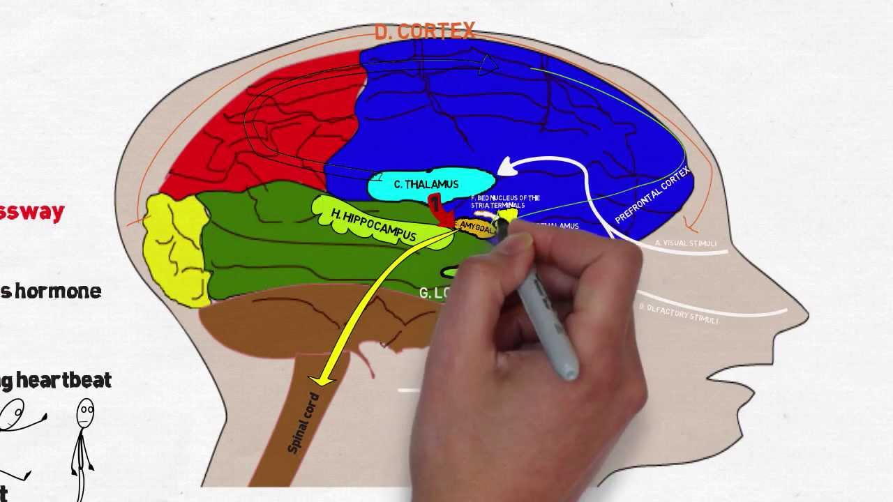
Schematic presentation of anxiety-related functional systems and brain structures. The arrows indicate the neural projections between two brain regions. The pathological interactions of the six critical regions in the anxiety network bring about functional abnormality for patients with anxiety disorders. Amyg, Amygdala BNST, Bed nucleus of the stria terminalis Hipp, Hippocampus Hyp, Hypothalamus PFC, Prefrontal cortex Str, Striatum.
The Brain Stem Relays Signals Between The Brain And Spinal Cord And Manages Basic Involuntary Functions
The brain stem connects the spinal cord to the higher-thinking centers of the brain. It consists of three structures: the medulla oblongata, the pons, and the midbrain. The medulla oblongata is continuous with the spinal cord and connects to the pons above. Both the medulla and the pons are considered part of the hindbrain. The midbrain, or mesencephalon, connects the pons to the diencephalon and forebrain. Besides relaying sensory and motor signals, the structures of the brain stem direct involuntary functions. The pons helps control breathing rhythms. The medulla handles respiration, digestion, and circulation, and reflexes such as swallowing, coughing, and sneezing. The midbrain contributes to motor control, vision, and hearing, as well as vision- and hearing-related reflexes.
You May Like: What Part Of The Brain Controls Autonomic Functions
You May Like: Can You Faint From Anxiety
Anxiety Caused By Lifestyle Habits
Never underestimate the effect your lifestyle can have on your anxiety levels. For example, some research has shown that those that do not exercise are more prone to developing anxiety, because their body has unused energy, and because they start producing less stress-coping hormones.
Similarly, although diet does not traditionally cause significant anxiety, it can still play a role. Caffeine, for example, can exacerbate mild anxiety symptoms. Unhealthy foods can cause you to feel unhealthy, which in turn causes stress that leads to anxiety. Drug use is frequently a cause of anxiety.
This is why even with therapy and medicine, a change to lifestyle habits is often necessary.
For more information, consider the following topics to read:
- Anxiety and Television
- Anxiety and Dehydration
Brain Structures Associated With Anxiety Disorders

Previous studies have shown that the amygdala, prefrontal cortex, bed nucleus of the stria terminalis , hippocampus, striatum, anterior insula, anterior cingulate cortex and hypothalamus were closely related to anxiety disorders. For instance, the activation in the hippocampus, amygdala and anterior insula during negative emotion processing was enhanced in patients with anxiety disorders. In addition, abnormal thalamic volume is an indicator of social anxiety, especially in children and adolescents. The neural networks that regulate social vigilance mainly comprise the bed nucleus of the stria terminalis and prefrontal cortex. Patients with anxiety disorders usually show behavioural avoidance, and the avoidance neural network includes the dorsal anterior cingulate cortex, anterior insula, sublenticular extended amygdala and temporal pole. Notably, the functional connectivity between the amygdala and the anterior insula is associated with the degree of avoidance. These structures, which are closely related to anxiety, may play unique roles in the development of cognitive and emotional capabilities among adolescents.
You May Like: What Is Phobia Mean
Environmental Causes Of Anxiety Disorders
We have spent a lot of time on CalmClinic discussing the biological and genetic components of anxiety. But for most people, anxiety is going to be largely environmental. For some, it may be difficult to find the cause at all, because not all anxiety is as simple as pointing to a specific moment.
Even if you have a genetic predisposition to anxiety , your environment is going to play a key role in whether you develop an anxiety disorder. Most believe that environment plays a triggering role in anxiety disorders, and in some cases may cause anxiety disorders by themselves.
In this case, environment includes everything that is not genetic – every experience you have, every place you go, and everything youve been taught.
According to a study of monozygotic twins and dizygotic twins , monozygotic twins – who both share the same DNA – were twice as likely to develop anxiety disorders than fraternal twins, but in each of these cases their genetics did not guarantee an anxiety disorder, which indicates that environment still plays a role.
Its also strongly believed that people can develop anxiety disorders from the environment alone. This is supported by the idea that anxiety can be treated without any medicine or surgery, indicating that a great deal of mental health is forged by life experiences.
Common environmental causes of anxiety include:
What Part Of The Brain Controls Depth Perception
Specifically the part of the brain that does depth perception based on binocular vision. I have searched all over the web and have turned up nothing. Is it the same part of the brain that processes all images from the retina?
This Scientific American article states that there is a process involved:
Visual-image processing from the eye to the brain happens in stages. Rudimentary features such as the orientation of edges, direction of motion, color, and so on are extracted early on in areas called V1 and V2 before reaching the next stages in the visual-processing hierarchy for a progressively more refined analysis. This stage-by-stage description is a caricature many pathways go back from stage to stageallowing the brain to play a kind of 20-questions game to arrive at a solution after successive iterations.
This process is discussed further in this article with what is known as the Laminart model.
Also Check: Phobia Psychology Definition
Lobes Of The Brain And What They Control
Each brain hemisphere has four sections, called lobes: frontal, parietal, temporal and occipital. Each lobe controls specific functions.
- Frontal lobe. The largest lobe of the brain, located in the front of the head, the frontal lobe is involved in personality characteristics, decision-making and movement. Recognition of smell usually involves parts of the frontal lobe. The frontal lobe contains Brocas area, which is associated with speech ability.
- Parietal lobe. The middle part of the brain, the parietal lobe helps a person identify objects and understand spatial relationships . The parietal lobe is also involved in interpreting pain and touch in the body. The parietal lobe houses Wernickes area, which helps the brain understand spoken language.
- Occipital lobe. The occipital lobe is the back part of the brain that is involved with vision.
- Temporal lobe. The sides of the brain, temporal lobes are involved in short-term memory, speech, musical rhythm and some degree of smell recognition.
When To See A Doctor
Autonomic disorders can be serious. People who experience symptoms of an autonomic disorder should see a doctor for a full diagnosis.
Talking to a doctor is particularly important for people with diabetes or other conditions that can increase the likelihood of autonomic disorders.
To diagnose the cause of ANS symptoms, a doctor will first assess a persons medical history for risk factors.
A doctor may also request one or more of the following:
- Tests to detect orthostatic hypotension: A doctor may measure OH using a tilt-table test. In this test, a person lies on a bed that tilts their body at different angles while a machine records their heart rate and blood pressure.
- Electrocardiogram: This test measures electrical activity within the heart.
- Sweat test: This test assesses whether the sweat glands are functioning correctly. A doctor uses electrodes to stimulate the sweat glands and measures the volume of sweat they produce in response to the stimulus.
- Pupillary light reflex test: This test measures how sensitive the pupils are to changes in light.
Also Check: Prodromal Period Schizophrenia
What Is The Gray Matter And White Matter
Gray and white matter are two different regions of the central nervous system. In the brain, gray matter refers to the darker, outer portion, while white matter describes the lighter, inner section underneath. In the spinal cord, this order is reversed: The white matter is on the outside, and the gray matter sits within.
Gray matter is primarily composed of neuron somas , and white matter is mostly made of axons wrapped in myelin . The different composition of neuron parts is why the two appear as separate shades on certain scans.
Each region serves a different role. Gray matter is primarily responsible for processing and interpreting information, while white matter transmits that information to other parts of the nervous system.
The Chemical Imbalance Theory
According to chemical imbalance theories, panic disorder symptoms can be attributed to imbalances in naturally occurring chemical messengers in the brain, known as neurotransmitters. These help communicate information between nerve cells brain throughout the brain.
The human brain is thought to have hundreds of these different types of neurotransmitters, and chemical imbalance theories suggest that a person can become more susceptible to developing panic disorder symptoms if one or more of these neurotransmitters do not remain balanced.
The neurotransmitters serotonin, dopamine, norepinephrine, and gamma-aminobutyric acid are specifically believed to be linked to mood and anxiety disorders. These neurotransmitters are in charge of regulating various bodily and mental functions.
First, serotonin is a neurotransmitter that is largely associated with mood, sleep, appetite, and other regulatory functions in the body. Experts have also found that reduced levels of serotonin are connected to depression and anxiety.
The neurotransmitter dopamine may also contribute to symptoms. Dopamine influences, among other functions, a persons energy levels, attention, rewards, and movement.
Norepinephrine is also related to anxiety as it released in the fight-or-flight response, or they physiological response to stress. Last, GABA plays a role in balancing excitement or agitation and inducing feelings of calm and relaxation through its inhibitory effect.
Don’t Miss: When Does Schizophrenia Develop In Males
The Coordinated Balance System
The human balance system involves a complex set of sensorimotor-control systems. Its interlacing feedback mechanisms can be disrupted by damage to one or more components through injury, disease, or the aging process. Impaired balance can be accompanied by other symptoms such as dizziness, vertigo, vision problems, nausea, fatigue, and concentration difficulties.
The complexity of the human balance system creates challenges in diagnosing and treating the underlying cause of imbalance. The crucial integration of information obtained through the vestibular, visual, and proprioceptive systems means that disorders affecting an individual system can markedly disrupt a persons normal sense of balance. Vestibular dysfunction as a cause of imbalance offers a particularly intricate challenge because of the vestibular systems interaction with cognitive functioning,2 and the degree of influence it has on the control of eye movements and posture.
Where Do Emotions Come From

The limbic system is a group of interconnected structures located deep within the brain. Its the part of the brain thats responsible for behavioral and emotional responses.
Scientists havent reached an agreement about the full list of structures that make up the limbic system, but the following structures are generally accepted as part of the group:
- Hypothalamus. In addition to controlling emotional responses, the hypothalamus is also involved in sexual responses, hormone release, and regulating body temperature.
- Hippocampus. The hippocampus helps preserve and retrieve memories. It also plays a role in how you understand the spatial dimensions of your environment.
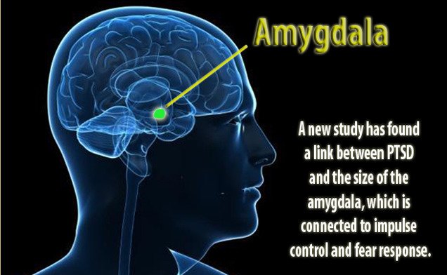
- Amygdala. The amygdala helps coordinate responses to things in your environment, especially those that trigger an emotional response. This structure plays an important role in fear and anger.
- Limbic cortex. This part contains two structures, the cingulate gyrus and the parahippocampal gyrus. Together, they impact mood, motivation, and judgement.
Recommended Reading: Apiphobic
Bed Nucleus Of The Stria Terminalis
The BNST is a component of the extended amygdala, which plays a critical role in the integration of autonomic and behavioral responses to stress . Earlier studies suggested that the CeA may be involved in processing explicit cue information associated with fear, while less explicit information associated with anxiety may activate the BNST . As stated earlier, given the vast evidence suggesting a role for both areas in anxiety, this view has begun to be contested .
Several studies have tested the effects of overall BNST modulation of different neurotransmitters in anxiety and in anxiety induced by acute or chronic stress. Changes in anxiety levels can be induced by intraBNST manipulation of CRF , glutamatergic AMPA receptors , serotonin 1A , calcium channel blockers , GABA synthesis , calcitonin generelated peptide , orexin A and B , and noradrenergic activity .
In awake animals, pharmacological activation of BNST elicits a rapid pressor response followed by bradycardia , whereas inactivation exacerbates restraintinduced increases in heart rate . This indicates that BNST signaling is necessary for inhibiting cardiovascular responses to stress. Modulation of heart rate by BNST stimulation or inhibition seems to be mediated by the parasympathetic nervous system .
Chronic stress and chronic corticosterone both increase BNST volume and modulation of cholinergic activity and galaninmediated signaling in the BNST can block the anxiogenic effects of restraint stress .
Effects Of Chronic Stress On Amygdalar Function And Anxiety
A large bulk of evidence suggests that chronic stress and early exposure to stress not only induce persistent anxiety even after 21 days from the end of stress , but also induce a series of functional and morphological changes in the brain. In the amygdala, changes in neural plasticity and electrophysiological responses including suppression of gamma-Aminobutyric acid currents , as well as a persistent increase in dendritic arborization of spiny neurons and amygdalar hypertrophy, have been reported as a result of chronic stress, effects are not restored by ceasing the stress , but can be restored with short environmental enrichment . Interestingly, some of these changes may be prevented by intraBLA antagonism of corticotrophin releasing factor , which is a critical hypothalamic activator of the HPA axis and mediator of stress responses in the brain.
Other brain areas involved in anxiety have also been reported to suffer changes and even damage as a result of chronic stress . For example, in animal models of chronic stress, the hippocampus and prefrontal cortex show atrophy, which may lead to memory impairments, whereas the amygdala hypertrophy may lead to increased anxiety and aggression .
You May Like: Very Late-onset Schizophrenia
Connections Between The Amygdala And Other Brain Regions Associated With Anxiety
The recent development of optogenetic approaches allowing the activation or inhibition of specific cell types or neuronal projections using the inducible expression of channel rhodopsins has begun to allow a much greater understanding of the circuitries associated with anxiety. In a recent study, it was shown that inhibition of vHPC input to the mPFC and bilateral, but not unilateral inhibition of the input to the BLA disrupts anxiety . In the same line, the activation of BLA inputs to the mPFC produces anxiogenic effects in the EPM and openfield tests, whereas inhibition of the structure produces anxiolytic effects . Furthermore, systemic activation of Kapa opiod receptors was shown to inhibit glutamate release from BLA projections to the BNST and prevent the anxiolytic effects induced by optogenetic activation of BLABNST projections, while deletion Kapa opiod receptors from amygdala neurons induces anxiolytic effects . In yet another study, it was reported that the stimulation of VTAprojecting BNST glutamatergic neurons has anxiogenic effects, while the activation of VTAprojecting BNST GABAergic neurons has anxiolytic effects, similar to the effects of direct inhibition of VTA GABAergic neurons .
Figure 2.
How Do Eyes Work
The images we see are made up of light reflected from the objects we look at. This light enters the eye through the cornea, which acts like a window at the front of the eye. The amount of light entering the eye is controlled by the pupil, which is surrounded by the iris the coloured part of the eye.
Because the front part of the eye is curved, it bends the light, creating an upside down image on the retina. The brain eventually turns the image the right way up.
The retina is a complex part of the eye, and its job is to turn light into signals about images that the brain can understand. Only the very back of it is light sensitive: this part of the retina is roughly the area of a 10p coin, and is packed with photosensitive cells called rods and cones.
Cones are the cells responsible for daylight vision. There are three kinds, each responding to a different wavelength of light: red, green and blue. The cones enable us to see images in colour and detail. Rods are responsible for night vision. They are sensitive to light but not to colour. In darkness, the cones do not function at all.
Read Also: How To Get Rid Of Fungus In Brain
Recommended Reading: Anxiety Causes Fainting
Interactions Between Neurotransmitter And Neuromodulatory Systems In Anxiety
It is critical to think of the brain as a dynamic system that is always changing and compensating to keep a balanced state, which may change over time with ageing, or chronic stress and could eventually lead to a pathological allostatic balance.
When we think of a neurotransmitter, we have the tendency to think of an individual synapse receiving a particular neurotransmitter and expressing receptors for this neurotransmitter only. In fact, the picture is much more complex, as a single neuron in cortex may participate in more than 10,000 synapses and receive different neurotransmitters, from glutamate, GABA, serotonin, or dopamine, to different peptides like orexins or neuropeptide Y. It may also respond to endocannabinoids, signaling fatty acids or microRNAs. In addition, released neurotransmitters can activate receptors located at the plasmatic membrane of neurons outside synapses. Moreover, astrocytes may also release their own transmitters onto synapses, including glutamate, dserine, and glycine, which are required for normal synaptic transmission and synaptic plasticity .
There are many other relevant interactions between stressmediators. Chronic CORT treatment mimicking chronic stress induces an increase in mRNA and protein levels of NE transporter and dopamine hydroxylase in the locus coeruleus, amygdala and hippocampus, suggesting increased NE synthesis .